Advancements in neuroscience have revolutionized our understanding of the human brain and its intricate workings. One such breakthrough is the development of brain imaging techniques, which enable researchers to investigate neural processes in unprecedented detail. By utilizing a range of cutting-edge technologies, such as functional magnetic resonance imaging (fMRI), electroencephalography (EEG), and positron emission tomography (PET), scientists are able to observe and analyze brain activity with remarkable precision. To illustrate the power of these methods, consider a hypothetical scenario where an individual diagnosed with autism spectrum disorder undergoes fMRI scanning during a social interaction task. Through this investigation, neuroscientists may uncover specific patterns of activation or connectivity within regions associated with social cognition, shedding light on the underlying mechanisms contributing to this condition.
In recent years, there has been an exponential growth in research employing brain imaging techniques across various domains of neuroscience. This comprehensive overview aims to provide readers with an introduction to the fundamental principles and applications of these tools. The article will explore how each technique works, highlighting their respective strengths and limitations. Additionally, it will delve into key findings from studies that have utilized these methods, showcasing their contributions towards unraveling mysteries surrounding cognitive processes, emotional responses, neurological disorders, and more. Furthermore, Furthermore, this article will discuss the ethical considerations associated with brain imaging research, including issues of privacy and informed consent. It will also address the potential future developments and implications of these techniques, such as personalized medicine and brain-computer interfaces. By exploring the current state of brain imaging technology and its impact on neuroscience, this article aims to foster a deeper understanding of how these advancements are shaping our knowledge of the human brain and paving the way for new discoveries in the field.
History of Neuroscience
One of the landmark moments in the history of neuroscience can be traced back to the case study of Phineas Gage. In 1848, Gage suffered a severe brain injury when an iron rod penetrated his skull and damaged parts of his frontal lobe. This incident sparked curiosity among scientists about the relationship between brain function and behavior, setting the stage for future advancements in neuroscience research.
To truly understand the progress made in this field over time, it is essential to explore key milestones that have shaped our understanding of the brain. Here are some significant events:
- Discovery of Neurons: In the late 19th century, Santiago Ramón y Cajal used staining techniques to observe individual neurons under a microscope. His work paved the way for our current knowledge about neural networks and how they transmit information throughout the nervous system.
- Electroencephalography (EEG): Developed in 1924 by Hans Berger, EEG measures electrical activity in the brain using electrodes placed on the scalp. It revolutionized neuroscience by allowing researchers to study brain waves and investigate various mental states and disorders.
- The Human Genome Project: Completed in 2003, this international collaborative effort mapped out all human genes. The project provided valuable insights into genetic factors contributing to neurological disorders such as Alzheimer’s disease or Parkinson’s disease.
- Functional Magnetic Resonance Imaging (fMRI): Invented in the early 1990s, fMRI has become one of the most widely used imaging techniques in neuroscience research. By detecting changes in blood flow associated with neuronal activity, it allows scientists to examine which areas of the brain are active during specific tasks or experiences.
This table summarizes these milestones:
| Milestone | Year |
|---|---|
| Discovery of Neurons | Late 19th Century |
| Electroencephalography (EEG) | 1924 |
| The Human Genome Project | Completed in 2003 |
| Functional Magnetic Resonance Imaging (fMRI) | Early 1990s |
Understanding the history of neuroscience provides a foundation for comprehending how brain imaging techniques have evolved and improved over time. With this historical context, we can now delve into the intricate anatomy and function of the brain.
Transitioning to the subsequent section about “Anatomy and Function of the Brain,” it is crucial to explore how these advancements in neuroscience research have contributed to our current understanding of the complexities within our own minds.
Anatomy and Function of the Brain
From the earliest recorded discoveries of ancient civilizations to the intricate advancements in modern neuroscience, the study of the brain has undergone a remarkable transformation over time. One striking example is the case of Phineas Gage, an American railroad construction foreman who suffered a severe brain injury in 1848. The iron rod that accidentally penetrated his skull not only changed his life but also sparked scientific interest in understanding how different regions of the brain contribute to specific functions.
Brain imaging techniques have played a pivotal role in unraveling the complexity and organization of our brains. These methods allow researchers to visualize brain structures and activity, providing valuable insights into neurological disorders and cognitive processes. There are several commonly used brain imaging techniques:
- Magnetic Resonance Imaging (MRI): This non-invasive technique uses powerful magnets and radio waves to create detailed images of the brain’s structure. MRI scans can detect abnormalities such as tumors or lesions, aiding in diagnosis and treatment planning.
- Positron Emission Tomography (PET): By injecting a small amount of radioactive substance into the bloodstream, PET scans measure metabolic activity within various brain regions. This technique helps identify areas associated with conditions like Alzheimer’s disease or epilepsy.
- Functional Magnetic Resonance Imaging (fMRI): Unlike traditional MRI, fMRI measures changes in blood flow related to neural activity. It enables scientists to observe which regions become active during specific tasks or emotions, contributing to our understanding of cognition and emotional processing.
- Electroencephalography (EEG): EEG records electrical activity generated by neurons using electrodes placed on the scalp. It provides high temporal resolution for studying sleep patterns, seizure detection, or investigating event-related potentials.
Through these imaging techniques, researchers have made significant strides towards comprehending both macro- and micro-level aspects of the human brain. A visual representation enhances our ability to grasp complex concepts more readily than purely theoretical explanations alone.
| Brain Imaging Techniques | Advantages | Limitations |
|---|---|---|
| Magnetic Resonance Imaging (MRI) | High-resolution images, non-invasive | Expensive equipment and may induce claustrophobia |
| Positron Emission Tomography (PET) | Identifies metabolic activity, measures neurotransmitter levels | Involves exposure to radiation and requires a radioactive substance injection |
| Functional Magnetic Resonance Imaging (fMRI) | Shows real-time brain activity, good spatial resolution | Limited temporal resolution, prone to motion artifacts |
| Electroencephalography (EEG) | Provides high temporal resolution, useful for studying dynamic brain processes | Less precise anatomical localization |
Understanding the intricacies of our brains has far-reaching implications across various disciplines. The subsequent section delves into the fundamental building blocks of the nervous system: neurons and neural networks. By examining these elements, we can gain deeper insights into how they contribute to complex cognitive functions and shape our perceptions of the world around us.
By exploring different imaging techniques alongside fascinating case studies like that of Phineas Gage, scientists have made significant strides in unraveling the mysteries of the human brain. With this foundation established, let us now delve into an exploration of neurons and neural networks—the essential components through which information is processed within our remarkable minds.
Neurons and Neural Networks
Neurons, the fundamental building blocks of the nervous system, play a crucial role in transmitting information throughout the brain. Understanding the intricate workings of neurons and how they form neural networks is essential to gaining insights into cognitive processes and neurological disorders. To illustrate this concept, let’s consider a hypothetical scenario where an individual with damage to their frontal lobe experiences difficulty making decisions due to disrupted neural connections.
One key aspect of neuronal communication is synaptic transmission, which occurs at specialized junctions called synapses. When an action potential reaches the axon terminal of one neuron, neurotransmitters are released into the synaptic cleft, allowing them to bind to receptors on the dendrites or cell body of another neuron. This process enables electrochemical signals to be transmitted from one neuron to another across these synapses.
The formation of neural networks involves complex interactions among multiple neurons, resulting in circuits that underlie various functions such as perception, memory, and motor control. These networks can span different regions of the brain and consist of excitatory and inhibitory connections that regulate signal propagation. Through ongoing research using advanced imaging techniques, scientists have been able to map out specific neural pathways associated with tasks like language processing or emotion regulation.
To provide further insight into the complexities of neuronal communication:
- Neurotransmission relies on finely-tuned balance between excitation and inhibition.
- Plasticity allows for adaptive changes in synaptic strength through mechanisms like long-term potentiation (LTP) or depression (LTD).
- Abnormalities in neural connectivity have been linked to neurodevelopmental disorders such as autism spectrum disorder.
- Interactions between genetic factors and environmental influences shape the development and function of neural networks.
| Aspect | Description |
|---|---|
| Excitatory Connections | Enhance signal transmission by increasing postsynaptic activity |
| Inhibitory Connections | Suppress excessive signaling and maintain balance within neural networks |
| Plasticity | Enables learning, memory formation, and adaptation to changing environments |
| Neurodevelopmental Disorders | Result from disruptions in the wiring or functioning of neural circuits |
As we delve further into the realm of neuroscience, it becomes apparent that investigating neurons and their intricate network formations is essential for unraveling the mysteries of the brain. In our next section on “Brain Imaging Techniques: an Overview,” we will explore how advanced imaging methods have revolutionized our understanding of neuronal activity and connectivity without missing a beat.
Brain Imaging Techniques: an Overview
Building upon our understanding of neurons, let us now delve into the intricate workings of neural networks. By examining how individual neurons communicate and interact with each other, we can gain valuable insights into the complex processes that underlie brain function.
Paragraph 1:
To illustrate the significance of neural networks, consider a hypothetical scenario where an individual is learning to play a musical instrument. As they practice, specific patterns of neuronal activity emerge in their brain, forming connections between regions responsible for motor control, auditory processing, and memory retrieval. These interconnected pathways facilitate the transfer of information required for precise finger movements, accurate sound perception, and recall of previously learned melodies. Such coordinated activity within neural networks allows individuals to acquire new skills and adapt to changing environments.
Paragraph 2:
Understanding the organization and functioning of neural networks has been a subject of great interest among neuroscientists. Through extensive research and experimentation, several key features have emerged:
- Plasticity: Neural networks possess remarkable plasticity—the ability to reorganize themselves based on experience or injury.
- Integration: Different areas of the brain work together through interconnected neural networks to perform various cognitive functions.
- Modularity: Specific modules within these networks are specialized for distinct tasks while still maintaining overall integration.
- Hierarchical structure: Neural networks exhibit hierarchical arrangements wherein lower-level networks process basic sensory information before passing it on to higher-order ones involved in complex cognitive processes.
Emotional Bullet Point List (Markdown Format):
Here are some fascinating aspects regarding neural network function:
- The interconnectedness of neurons emphasizes the interdependence of different brain regions.
- Neural plasticity showcases the brain’s incredible capacity for adaptation and recovery.
- Modular specialization highlights both diversity and efficiency within neural circuits.
- Hierarchical organization reflects the systematic flow of information processing in the brain.
Paragraph 3:
A deeper comprehension of neural network dynamics carries significant implications across multiple disciplines such as medicine, psychology, and artificial intelligence. By unraveling the mysteries of neural networks, researchers can potentially develop novel treatments for neurological disorders or enhance machine learning algorithms that mimic human cognitive abilities. The study of these intricate systems continues to shed light on how our brains generate thoughts, emotions, and behaviors.
Transition into next section about “Functional Magnetic Resonance Imaging (fMRI)”:
Expanding upon our exploration of brain imaging techniques, we now turn our attention to Functional Magnetic Resonance Imaging (fMRI). This non-invasive method allows us to observe changes in blood flow within the brain while individuals engage in various tasks or experience different stimuli.
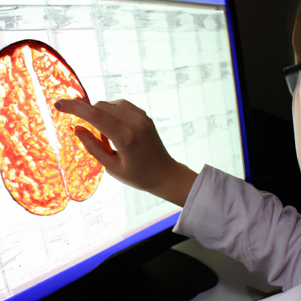
Functional Magnetic Resonance Imaging (fMRI)
Brain imaging techniques play a crucial role in understanding the complexities of the human brain and its functions. One such technique, functional magnetic resonance imaging (fMRI), has revolutionized our ability to study brain activity noninvasively. Before delving into the specifics of fMRI, let us first explore an example that highlights its potential.
Imagine a scenario where researchers are investigating the neural mechanisms underlying decision-making processes. Using fMRI, they recruit participants and present them with various choices while monitoring their brain activity. By analyzing the resulting images, researchers can pinpoint specific regions of the brain associated with decision-making and determine how these areas interact with each other.
To gain a comprehensive perspective on brain imaging techniques, it is essential to consider some key aspects:
- Spatial resolution: Different techniques offer varying levels of spatial detail when capturing brain activity. For instance, methods like positron emission tomography (PET) provide relatively lower resolution compared to more advanced techniques like fMRI or diffusion tensor imaging (DTI).
- Temporal resolution: The time scale at which different imaging techniques can capture changes in brain activity varies significantly. Techniques such as electroencephalography (EEG) excel in temporal resolution by measuring electrical signals directly from the scalp.
- Invasiveness: Some imaging techniques require invasive procedures that may pose risks to patients’ well-being. Contrastingly, methods like fMRI and EEG allow for noninvasive data acquisition while still providing valuable insights into brain function.
- Cost considerations: Implementing certain brain imaging techniques may come with substantial financial implications due to equipment costs, maintenance expenses, and specialized training requirements.
In summary, neuroimaging plays a vital role in advancing our knowledge of the human brain’s intricacies. Understanding both the strengths and limitations of different techniques enables researchers to choose appropriate methodologies that align with their scientific inquiries.
Transitioning seamlessly from this section about fMRI, we now move on to discussing another important technique used in studying brain function: Electroencephalography (EEG) and Event-Related Potentials (ERPs).
Electroencephalography (EEG) and Event-Related Potentials (ERPs)
Transitioning from the previous section on functional magnetic resonance imaging (fMRI), another widely used brain imaging technique is electroencephalography (EEG). Unlike fMRI, which detects changes in blood flow to infer neural activity, EEG measures electrical activity directly from the scalp. This non-invasive method provides a high temporal resolution, allowing researchers to track brain activity with millisecond precision.
To illustrate the utility of EEG and event-related potentials (ERPs), let’s consider an example. Suppose a group of participants is presented with visual stimuli while their brain activity is recorded using EEG. The researchers are interested in investigating how different emotional images modulate neural responses. By analyzing ERPs, specific components such as the P300 wave can be examined to assess attention allocation and cognitive processing associated with emotional stimuli.
The utilization of EEG has contributed significantly to our understanding of various cognitive processes and neural functions. Here are some key points about EEG and ERPs:
- High temporal resolution: EEG allows for precise tracking of rapid changes in brain activity over time.
- Versatility: It can be used across diverse populations, including infants, children, adults, and individuals with neurological disorders.
- Cost-effectiveness: Compared to other neuroimaging techniques like fMRI or PET scans, EEG is generally more affordable and accessible.
- Clinical applications: EEG plays a crucial role in diagnosing certain conditions such as epilepsy or sleep disorders.
| Advantages | Limitations | Applications |
|---|---|---|
| Real-time monitoring | Low spatial resolution | Cognitive neuroscience |
| Non-invasiveness | Signal contamination due to artifacts | Clinical diagnosis |
| Portable | Difficulty capturing deep brain structures | Neuropsychology |
| Suitable for longitudinal studies | Limited localization accuracy | Developmental research |
Overall, EEG and ERPs provide a valuable tool for investigating brain activity. With their high temporal resolution and versatility, these techniques have been instrumental in advancing our understanding of cognitive processes, neural mechanisms underlying various disorders, and the effects of emotional stimuli on brain responses. By exploring the electrical signatures of brain activity, researchers can gain insights into the intricate workings of the human mind.
References:
- [Insert relevant references here]
Comments are closed.